Treatment · lumbar
Microdiscectomy: How It Works, Recovery & Risks
Microdiscectomy is a microscope-assisted operation that removes the part of a herniated lumbar disc pressing on a nerve root, relieving sciatica while preserving most of the disc.
Microdiscectomy is one of the most established operations for a herniated lumbar disc that is pressing on a nerve root and causing sciatica. Using an operating microscope, the surgeon removes the small fragment compressing the nerve through a modest incision, leaving most of the disc and surrounding structures intact. This page sets out, in neutral terms, what the procedure involves, who may benefit, how it compares with other approaches, and the realistic balance of benefits and risks.
What is microdiscectomy?
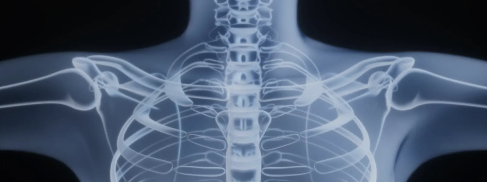
The discs between the vertebrae cushion the spine and allow movement. When the soft centre of a disc pushes through its outer wall, the resulting fragment can press on a nearby nerve root in the lower back. This commonly causes pain that radiates down the leg, often with numbness, tingling or weakness — the pattern known as sciatica.
Microdiscectomy, also called micro-lumbar discectomy, removes the herniated fragment using an operating microscope for a magnified, well-lit view. The microscope allows the surgeon to work through a smaller incision than older open techniques while still seeing the nerve clearly. Only the protruding fragment and any loose pieces likely to herniate again are removed; the rest of the disc is preserved. The aim is to relieve pressure on the nerve, so the target is nerve-related leg pain rather than generalised back pain.
Who is a candidate?
Microdiscectomy is generally considered for people who have:
- Leg pain, numbness or weakness in a pattern that matches a disc herniation seen on MRI.
- Symptoms that have not settled after a reasonable period of non-surgical care, such as physiotherapy, activity modification, medication or spinal injections.
- Pain that significantly limits daily activities, work or sleep.
- Progressive nerve-related weakness, which may prompt earlier consideration of surgery.
It is usually less appropriate when the main complaint is back pain without nerve compression, when there is significant spinal instability that might need a different operation, or when imaging does not match the symptoms. Sudden severe weakness, or loss of bladder or bowel control, are warning signs that need urgent assessment rather than planned surgery.
How the procedure is performed
The general steps, which may vary by centre, are:
- Anaesthesia and positioning. The operation is usually carried out under general anaesthesia, with the patient positioned face down.
- Confirming the level. X-ray imaging is used to identify and confirm the correct disc level.
- The incision. A small incision is made in the midline of the lower back over the affected level.
- Reaching the disc. The muscles are gently moved aside and a small amount of bone or ligament may be removed to expose the nerve and the herniation.
- Removing the fragment. Under the microscope, the nerve is carefully protected and the herniated fragment is removed, freeing the nerve.
- Checking and closure. The surgeon confirms the nerve moves freely, then closes the incision in layers.
Minimally invasive vs open
Microdiscectomy is itself a refinement of open discectomy: the microscope allows a smaller wound and a clearer view, so it is often described as minimally invasive compared with traditional open surgery. Other minimally invasive options, such as tubular discectomy or endoscopic discectomy, use tubes or a camera to reduce muscle disruption further.
Each approach has trade-offs. A more open exposure may be preferred for complex anatomy, large or migrated fragments, or revision surgery, while narrower approaches may reduce early tissue trauma in suitable cases. For most uncomplicated lumbar herniations, the published results of these techniques are broadly comparable, and the choice is individualised rather than one-size-fits-all.
Benefits and risks
Potential benefits include reliable relief of nerve-related leg pain in well-selected patients, a relatively small incision, early mobilisation and, often, day-case or short-stay treatment. It is one of the most studied spinal operations, with a long track record.
Risks, though uncommon, are real and include:
- Recurrent herniation at the same disc level.
- Dural tear with leakage of spinal fluid.
- Nerve irritation or, rarely, injury.
- Infection or bleeding.
- Persistent or incompletely relieved symptoms.
- The possibility of further surgery.
Because the operation targets the pinched nerve, it is not designed to cure mechanical back pain, and some back discomfort may remain. A clear discussion about the expected benefit in your own case is central to informed consent.
Alternatives
Many disc herniations improve over weeks to months without surgery. Non-surgical options include activity modification, physiotherapy, pain-relieving medication and spinal injections that can reduce inflammation around the nerve. Where surgery is indicated, alternatives to microdiscectomy include endoscopic or tubular discectomy and, less commonly, open discectomy. If there is associated instability or recurrent problems, a fusion procedure may occasionally be considered. The right path depends on the diagnosis, the severity and duration of symptoms, and your preferences after a full discussion with your surgeon.
Recovery and outlook
Most patients walk on the day of surgery and go home the same day or the next. In the first few weeks it is usual to avoid heavy lifting, repeated bending and twisting, and prolonged sitting, while steadily increasing gentle walking and daily activity. Physiotherapy often guides a graded return to work and exercise, sooner for sedentary roles and later for heavy manual work.
Leg pain frequently improves soon after surgery, while numbness or weakness that has been present for some time may take longer to settle and occasionally does not fully resolve. Recovery depends on individual factors such as how long the nerve was compressed and general health, so a particular outcome cannot be guaranteed.
When to seek a specialist opinion
Consider a specialist opinion if you have persistent leg pain, numbness or weakness in a nerve pattern that has not improved with non-surgical care, or if symptoms are disrupting daily life, work or sleep. Seek urgent medical attention for sudden severe leg weakness, loss of bladder or bowel control, or numbness in the saddle area. A spine specialist can match your symptoms to your imaging and advise whether microdiscectomy, another procedure or continued non-surgical care is most appropriate for you.
Frequently asked questions
What is the difference between microdiscectomy and discectomy?
Discectomy is the general term for removing herniated disc material. Microdiscectomy refers to doing this with the help of an operating microscope through a small incision, which gives magnification and better lighting and allows a smaller wound than older open techniques.
Is the whole disc removed?
No. Only the herniated fragment pressing on the nerve, and any loose pieces within the disc that could cause early recurrence, are removed. The bulk of the disc is preserved so it can continue to act as a spacer.
Will microdiscectomy cure my back pain?
Microdiscectomy is designed mainly to relieve leg pain (sciatica) from a pinched nerve. It is less reliable for mechanical back pain, and some back pain may persist. Setting realistic expectations is an important part of planning.
How long does recovery take?
Many people walk on the day of surgery and go home the same day or the next. A graded return to normal activity usually takes a few weeks, with heavy manual work and high-impact exercise resumed later on your surgeon’s advice.
Can the disc herniate again after surgery?
Yes. Because most of the disc is preserved, a recurrent herniation can occur at the same level. This is uncommon but recognised, and a minority of patients may need further surgery.
What are the risks of microdiscectomy?
Recognised risks include recurrence, dural tear with spinal-fluid leak, nerve irritation or injury, infection, bleeding, and the small chance of persistent symptoms or further surgery. Your surgeon will discuss how these apply to you.
How is it different from endoscopic discectomy?
Both remove the offending fragment. Microdiscectomy uses a microscope through a small open incision, while endoscopic discectomy uses a camera passed through a narrow tube. The best option depends on the herniation and the surgeon’s assessment; outcomes for suitable patients are broadly similar.
Do I definitely need surgery for a slipped disc?
Often not. Many disc herniations settle with time and non-surgical care. Surgery is usually considered when severe or persistent nerve pain does not improve, or when there is significant or progressive weakness. A specialist can advise on timing.