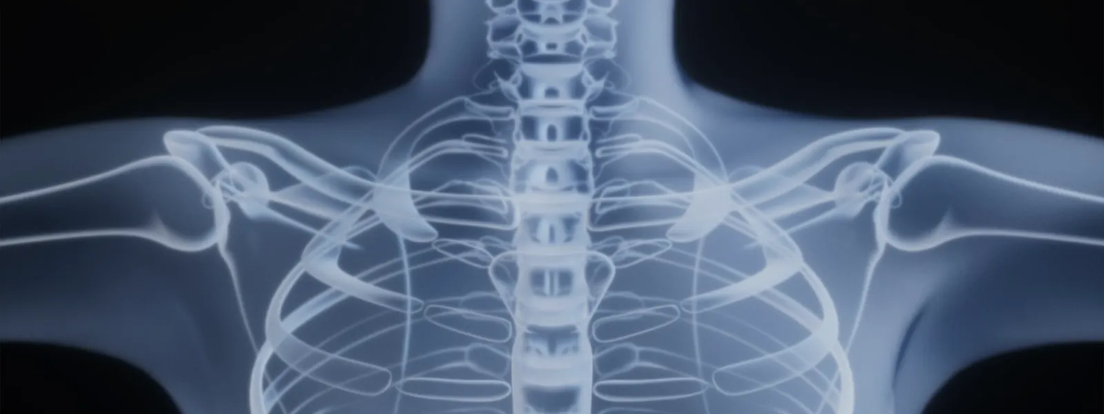
Treatment · lumbar
Micro-Endoscopic Decompression Explained
Micro-endoscopic decompression (MED) uses a small tubular retractor and an endoscope to relieve pressure on the nerves of the lower spine from stenosis or a disc, through a small incision.
Micro-endoscopic decompression is a minimally invasive way to relieve pressure on the nerves of the lower spine through a small incision. Using a narrow tubular retractor and an endoscope, the surgeon can not only remove disc material but also widen a narrowed spinal canal or nerve exit, making it particularly useful for spinal stenosis. This page explains how the technique works, who it may help, and what recovery involves, as general education rather than personal medical advice.
What is micro-endoscopic decompression?
The term decompression means relieving pressure on the spinal nerves. In micro-endoscopic decompression (MED), this is done through a tubular system: a series of dilators gently spread the back muscles, and a small tube is left in place to provide a working channel. An endoscope inside the tube gives a magnified, illuminated view.
Through this channel, the surgeon can remove the structures crowding the nerves — which may include thickened ligament, bone spurs and overgrown bony edges in spinal stenosis, as well as herniated disc material. This broader capacity to address stenosis distinguishes it from a procedure aimed only at removing a disc fragment.
Who is a candidate? (Indications)
Micro-endoscopic decompression is generally considered when nerve compression in the lower back causes significant symptoms that have not settled with appropriate non-surgical care. Common indications include:
- Lumbar spinal stenosis causing leg pain, heaviness or numbness, especially when walking or standing.
- A slipped (herniated) disc pressing on a nerve root.
- Narrowing of the bony exit where a nerve leaves the canal.
- Symptoms that match the imaging findings and have not improved with conservative treatment.
It is most suitable where the spine is stable and the main problem is compression rather than instability. Where there is significant instability, a stabilising procedure may be required instead. Suitability is decided individually.
How the procedure is performed
- Anaesthesia and positioning. The procedure is performed under general or regional anaesthesia, with the patient face-down.
- Small incision. A small incision is made over the affected level under imaging guidance.
- Muscle dilation. Dilators gently spread the muscle fibres, and a tubular retractor is placed to create a working channel.
- Endoscopic view. The endoscope provides a clear, magnified image of the spinal structures.
- Decompression. Thickened ligament, bone and, where present, herniated disc material are removed to free the trapped nerves.
- Checking the result. The surgeon confirms that the nerves are adequately decompressed.
- Closure. The tube is removed and the small incision is closed with minimal stitching.
Benefits and risks
Possible benefits. Because the muscle is dilated rather than stripped, the technique aims to reduce blood loss, post-operative pain and hospital stay, and may allow early mobilisation. It can address stenosis as well as disc problems through a small approach.
Risks and limitations. As with any spinal surgery, there are real risks, including:
- Infection or wound problems.
- Bleeding.
- Dural tear with cerebrospinal fluid leak.
- Nerve irritation or injury, with possible weakness or numbness.
- Incomplete relief of symptoms.
- Recurrent disc herniation or recurrent narrowing over time.
- Rarely, the development of instability requiring further surgery.
The technique is demanding and works best in carefully selected patients. No procedure can guarantee complete relief.
Alternatives
Depending on the diagnosis, alternatives include microdiscectomy for a focal disc herniation, endoscopic discectomy, traditional open decompression, or continued non-surgical care such as physiotherapy, activity modification and spinal injections. Where instability is present, a decompression combined with fusion may be more appropriate. The right choice depends on the anatomy, the goals of treatment and the stability of the spine.
Recovery and outlook
Recovery is generally quicker than with open surgery, but remains individual.
- Early days: Many patients go home the same day or after a short stay and begin gentle walking.
- First weeks: Gradual return to light activity, with restrictions on heavy lifting, bending and twisting.
- Following weeks: Activity is steadily increased, often with physiotherapy, as symptoms settle.
Leg symptoms often improve relatively quickly, while back symptoms may respond less predictably. Outcomes vary with the underlying condition.
When to seek a specialist opinion
Consider a specialist assessment if you have persistent leg pain, numbness or walking difficulty from nerve compression that limits daily life despite appropriate non-surgical treatment. Seek urgent medical attention for new or worsening leg weakness, numbness around the groin or buttocks, or loss of bladder or bowel control, as these may indicate a serious problem. A spine specialist can advise whether micro-endoscopic decompression or another approach is most appropriate for you.
Frequently asked questions
How is this different from a microdiscectomy?
A microdiscectomy mainly removes herniated disc material pressing on a nerve. Micro-endoscopic decompression can do this but also widen a narrowed canal or nerve exit in spinal stenosis, removing bone and thickened ligament as well as disc.
Does it involve fusion?
No. A pure decompression relieves pressure on the nerves without joining vertebrae. If the spine is unstable, a separate stabilising procedure may be needed, which is a different operation.
What conditions does it treat?
It is commonly used for lumbar spinal stenosis and for a slipped (herniated) disc compressing a nerve, where the main goal is to free trapped nerves through a small approach.
How big is the incision?
The incision is small, typically allowing a narrow tubular retractor to pass through to the spine. Exact size depends on the system used and the anatomy being treated.
Will my leg pain improve?
Relieving pressure on the nerve often improves leg pain, numbness and walking difficulty. Back pain may improve less predictably, and outcomes vary between individuals.
What are the risks?
Risks include infection, bleeding, dural tear with fluid leak, nerve irritation or injury, incomplete relief, and the possibility of recurrent symptoms or the need for further surgery.
How long is recovery?
Many patients go home the same day or after a short stay and resume light activity within days to weeks. Heavier activity is reintroduced gradually under guidance.