Treatment · lumbar / cervical
Endoscopic Discectomy: How It Works, Recovery & Risks
Endoscopic discectomy is a keyhole spine operation that removes a herniated disc fragment pressing on a nerve, using a small tube and camera passed through a portal a few millimetres wide.
Endoscopic discectomy is one of the least invasive ways to remove a herniated disc fragment that is pressing on a spinal nerve. It uses a slim camera and instruments passed through a portal only a few millimetres wide, allowing the surgeon to reach the nerve and remove the offending fragment while disturbing very little of the surrounding muscle and bone. This page explains, in neutral terms, what the procedure involves, who it may suit, how it compares with other techniques, and the balance of benefits and risks.
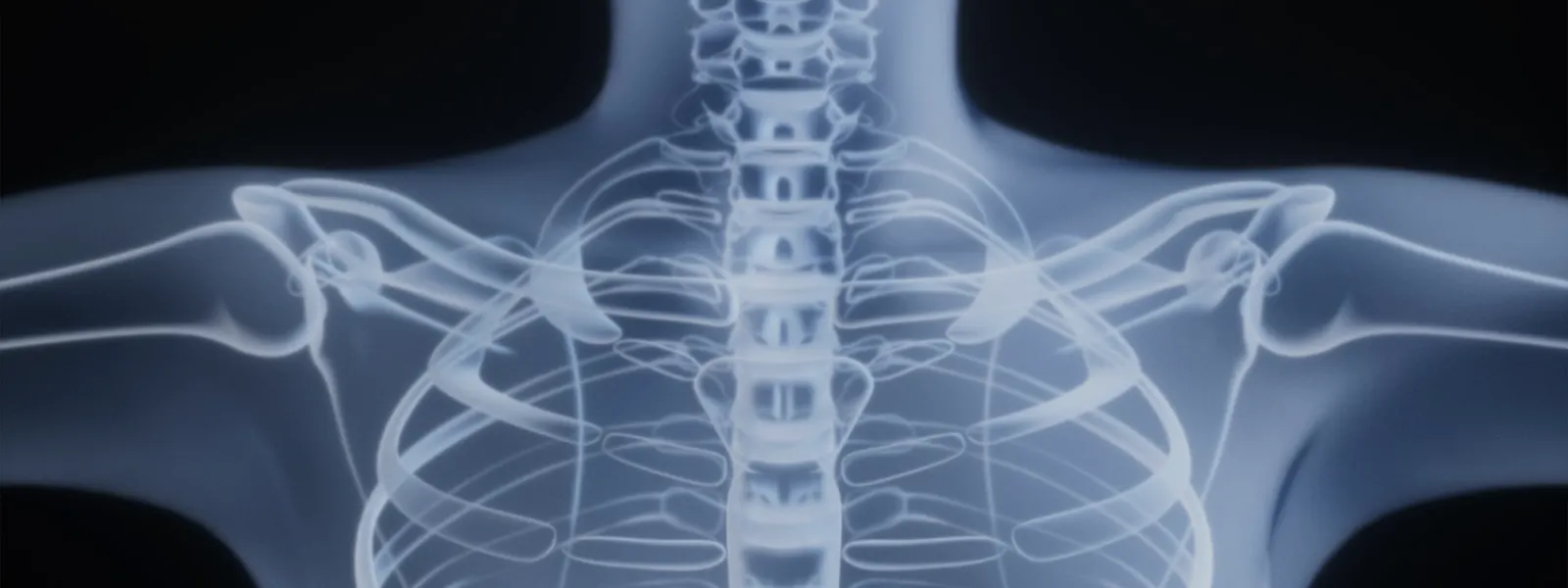
What is endoscopic discectomy?
The intervertebral discs sit between the vertebrae and act as cushions. When the soft inner part of a disc pushes through its tougher outer wall, the resulting bulge or fragment can press on a nearby nerve root. This often produces pain, numbness, tingling or weakness travelling along the path of that nerve — down the leg in the lumbar spine (sciatica) or into the arm in the cervical spine.
Endoscopic discectomy, sometimes called full-endoscopic discectomy, removes the protruding fragment using an endoscope: a narrow instrument carrying a camera, light and a channel for fine surgical tools. Continuous saline irrigation keeps the view clear. Because the working portal is so small, the technique is often described as keyhole spine surgery. The goal is targeted decompression of the nerve, not removal of the entire disc.
Who is a candidate?
Endoscopic discectomy is generally considered for people who have:
- Leg pain (sciatica) or arm pain caused by a confirmed disc herniation pressing on a nerve root.
- Symptoms that correspond to the level and side of the herniation seen on MRI.
- Pain, numbness or weakness that has not settled with a reasonable period of non-surgical care, such as activity modification, physiotherapy, medication or spinal injections.
- Symptoms that significantly affect daily life, or progressive nerve-related weakness.
It is usually less suitable when the main problem is mechanical back or neck pain without nerve compression, when there is significant spinal instability, or when the anatomy of the herniation makes endoscopic access difficult. Urgent features such as loss of bladder or bowel control, or rapidly worsening weakness, are warning signs that need immediate medical attention rather than routine planning.
How the procedure is performed
While details vary between centres and between the lumbar and cervical spine, the general steps are:
- Anaesthesia and positioning. The procedure may be done under local anaesthesia with sedation or under general anaesthesia. You are positioned to give the surgeon access to the affected level, usually face down for lumbar cases.
- Locating the level. Live X-ray (fluoroscopy) is used to confirm the correct disc level and plan the path of the portal.
- Creating the portal. A small skin incision is made and a guide is passed towards the disc. A working tube (cannula) is placed, through which the endoscope is introduced.
- Visualising the nerve. The camera projects a magnified view onto a screen. Saline irrigation keeps the field clear and helps control minor bleeding.
- Removing the fragment. Fine instruments remove the herniated fragment compressing the nerve. The surgeon confirms that the nerve is free and moves easily.
- Closure. The instruments are withdrawn and the small incision is closed, often with a single stitch or adhesive strip.
Minimally invasive vs open
Compared with traditional open surgery, endoscopic discectomy involves a smaller incision and less disruption of the muscles attached to the spine. Open discectomy, and the microscope-assisted microdiscectomy that has largely replaced it for many cases, provides a wider corridor and may be preferred for certain herniations, revision surgery or complex anatomy.
The endoscopic approach can mean less tissue trauma and, for some patients, a quicker early recovery, but it is technically demanding and is not suitable for every herniation. The decision between endoscopic, microscopic and open techniques is made case by case, weighing the position of the fragment, the patient’s anatomy and the evidence for each option. No single technique is best for everyone.
Benefits and risks
Potential benefits include a small incision, reduced damage to surrounding muscle, the possibility of day-case treatment and, for suitable patients, early mobilisation. Targeted removal of the fragment is intended to relieve nerve-related leg or arm pain.
As with any operation, there are risks. These include:
- Incomplete removal of the fragment or persistent symptoms.
- Recurrent herniation at the same level.
- Irritation or, rarely, injury to a nerve root.
- Dural tear with leakage of spinal fluid.
- Infection or bleeding.
- The possibility of needing further or open surgery.
Endoscopic discectomy mainly addresses nerve compression; it is not designed to relieve generalised back or neck pain, and expectations should reflect this. A frank discussion of the likely benefit in your particular case is an essential part of consent.
Alternatives
Not everyone with a disc herniation needs surgery. Many herniations improve over weeks to months with non-surgical care, including activity modification, physiotherapy, pain-relieving medication and, in some cases, spinal injections that can reduce inflammation around the nerve. Where surgery is appropriate, alternatives to the endoscopic approach include microdiscectomy and open discectomy. In a minority of cases with instability or recurrent problems, a fusion procedure may be considered instead. The most appropriate path depends on the diagnosis, the severity of symptoms and your preferences after a full discussion.
Recovery and outlook
Recovery varies from person to person. Many patients walk on the day of surgery and go home the same day or after a short stay. Early on, it is usual to avoid heavy lifting, repeated bending and twisting, and prolonged sitting, while gradually increasing gentle activity. A structured return to work and exercise — often guided by physiotherapy — typically unfolds over several weeks, sooner for sedentary roles and later for heavy manual work.
Relief of nerve-related pain is often noticed early, although numbness or weakness that has been present for a while can take longer to settle and may not fully resolve. Outcomes depend on factors such as how long the nerve has been compressed and individual healing, so it is not possible to guarantee a particular result.
When to seek a specialist opinion
It is reasonable to seek a specialist opinion if you have persistent leg or arm pain, numbness or weakness that follows a nerve pattern and has not improved with non-surgical care, or if symptoms are interfering with daily life. Seek urgent medical care for sudden severe weakness, loss of bladder or bowel control, or numbness in the saddle area, as these may indicate a problem needing prompt assessment. A spine specialist can correlate your symptoms with your imaging and explain whether endoscopic discectomy, another procedure or continued non-surgical care is the most appropriate option for you.
Frequently asked questions
Is endoscopic discectomy the same as microdiscectomy?
Both remove the herniated fragment compressing a nerve, but the access differs. Microdiscectomy uses an operating microscope through a small open incision, while endoscopic discectomy uses a camera passed down a narrow tube. Outcomes for suitable patients are broadly comparable; the right choice depends on the anatomy of the herniation and the surgeon’s assessment.
Will the whole disc be removed?
No. The aim is to remove only the loose or protruding fragment that is pressing on the nerve, preserving as much healthy disc as possible. The remaining disc continues to act as a spacer between the vertebrae.
How long does the operation take?
A single-level endoscopic discectomy commonly takes around 45 to 90 minutes, though this varies with the location of the herniation, the patient’s anatomy and whether one or more levels are treated.
Will I be awake during the procedure?
It can be performed under local anaesthesia with sedation, which allows some patients to give feedback during surgery, or under general anaesthesia. The choice depends on the level treated, your health and the preferences agreed with your anaesthetist and surgeon.
How soon can I return to work?
Many people with desk-based jobs return within one to three weeks, while those doing heavy manual work usually need longer. Recovery is individual, and your surgical team will give advice based on your job and progress.
Can the disc herniate again?
Yes. A recurrent herniation can occur at the same level because the remaining disc can protrude again. This is uncommon but recognised, and it is one reason follow-up and sensible activity advice matter.
What are the main risks?
Risks include incomplete removal of the fragment, recurrence, nerve irritation or injury, infection, bleeding, dural tear with leak of spinal fluid, and the small chance of needing further surgery. Your surgeon will discuss how these apply to your case.
How do I know if I am a suitable candidate?
Suitability depends on your symptoms, examination findings and MRI, and whether non-surgical care has been tried. A specialist assessment is needed to confirm that the herniation seen on imaging matches your symptoms before considering surgery.